Metabolic Phenotyping
The Metabolic Phenotyping Core serves as a pivotal resource for researchers seeking to characterize intricate metabolic phenotypes across diverse model systems and disease contexts. With its multitude of services that enable a comprehensive understanding of fuel utilization in cultured cells, tissue biopsies, and intact organisms, the core helps investigators unravel the elegant and nuanced complexities of metabolic regulation.
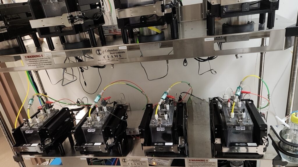
At the heart of the core’s capabilities lies the Oxymax Comprehensive Lab Animal Monitoringsystem (CLAMS), facilitating the precise determination of whole animal energy expenditure. Additionally, the core offers expertise in evaluating oxidative phosphorylation and glycolysis, employing the state-of-the-art Seahorse XF oxygen monitor to scrutinize mitochondrial functionin isolated organelles, intact cells, or tissues.
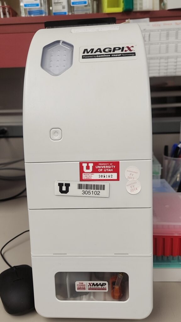
The core also boasts proficiency in assessing body composition in small animals through cutting-edge NMR technology. Beyond metabolic assessments, we excels in multiplex analytemeasurement, harnessing the power of instruments like the Multiplex reader Magpix to simultaneously scrutinize up to 50 analytes from serum, plasma, or other bodily fluids. In addition to this impressive instrument repertoire, the core provides essential support infrastructure, including refrigerated rodent incubators for studying animals across varying environmental conditions. Furthermore, an EPR system enables the detection of free radicals from both cells and tissues, enriching our understanding of altered electron exchange in the control of tissue function. Collaborative ventures with esteemed research groups further enhance our offerings, with specialized services such as euglycemic clamping conducted in partnership with Dr. Holland’s lab and high resolution respirometry utilizing the Oroboros system in collaboration with Dr. Funai’s lab.
Through these collaborative efforts and cutting-edge methodologies, the Metabolic Phenotyping Core stands poised to advance metabolic research and foster scientific discovery.
Service Rates
Requesting Services
Existing users may login directly to the Resource Scheduling System to schedule or order services. This system is cores-wide and uses University of Utah uNID authentication.


Hours of Operation
9:00 am to 5:00 pm
Monday - Friday
801-581-2425
Shipping Address
Metabolic Phenotyping Core
Radiobiology, Bldg 585
50 N 2030 E, Rm 151
Salt Lake City, UT 84112
Shipping Address
Large Instruments
Comparative Medicine Center - Loading Dock
50 N 2030 E
Salt Lake City, UT 84112
Recent Mentions
- Clark, A. M., Yu, D., Neiswanger, G., Zhu, D., Zou, J., Maschek, J. A., Burgoyne, T., & Yang, J. (2024). Disruption of CFAP418 interaction with lipids causes widespread abnormal membrane-associated cellular processes in retinal degenerations. JCI Insight, 9(1). https://doi.org/10.1172/jci.insight.162621
- Eberhardt, D. R., Lee, S. H., Yin, X., Balynas, A. M., Rekate, E. C., Kraiss, J. N., Lang, M. J., Walsh, M. A., Streiff, M. E., Corbin, A. C., Li, Y., Funai, K., Sachse, F. B., & Chaudhuri, D. (2022). EFHD1 ablation inhibits cardiac mitoflash activation and protects cardiomyocytes from ischemia. J Mol Cell Cardiol, 167, 1-14. https://doi.org/10.1016/j.yjmcc.2022.03.002
- Ferrara, P. J., Lang, M. J., Johnson, J. M., Watanabe, S., McLaughlin, K. L., Maschek, J. A., Verkerke, A. R. P., Siripoksup, P., Chaix, A., Cox, J. E., Fisher-Wellman, K. H., & Funai, K. (2023). Weight loss increases skeletal muscle mitochondrial energy efficiency in obese mice. Life Metab, 2(2). https://doi.org/10.1093/lifemeta/load014
- Johnson, J. M., Peterlin, A. D., Balderas, E., Sustarsic, E. G., Maschek, J. A., Lang, M. J., Jara-Ramos, A., Panic, V., Morgan, J. T., Villanueva, C. J., Sanchez, A., Rutter, J., Lodhi, I. J., Cox, J. E., Fisher-Wellman, K. H., Chaudhuri, D., Gerhart-Hines, Z., & Funai, K. (2023). Mitochondrial phosphatidylethanolamine modulates UCP1 to promote brown adipose thermogenesis. Sci Adv, 9(8), eade7864. https://doi.org/10.1126/sciadv.ade7864
- Li, Y., Talbot, C. L., Chandravanshi, B., Ksiazek, A., Sood, A., Chowdhury, K. H., Maschek, J. A., Cox, J., Babu, A. K. S., Paz, H. A., Babu, P. V. A., Meyerholz, D. K., Wankhade, U. D., Holland, W., Shyong Tai, E., Summers, S. A., & Chaurasia, B. (2022). Cordyceps inhibits ceramide biosynthesis and improves insulin resistance and hepatic steatosis. Sci Rep, 12(1), 7273. https://doi.org/10.1038/s41598-022-11219-3Nielson, J. R., Nath, A. K., Doane, K. P., Shi, X.,
- Nielson, J. R., Nath, A. K., Doane, K. P., Shi, X., Lee, J., Tippetts, E. G., Saha, K., Morningstar, J., Hicks, K. G., Chan, A., Zhao, Y., Kelly, A., Hendry-Hofer, T. B., Witeof, A., Sips, P. Y., Mahon, S., Bebarta, V. S., Davisson, V. J., Boss, G. R., . . . Peterson, R. T. (2022). Glyoxylate protects against cyanide toxicity through metabolic modulation. Sci Rep,12(1), 4982. https://doi.org/10.1038/s41598-022-08803-y
Citing Our Facility
Acknowledgments
We would like to thank you for acknowledging the our facility. This recognition allows us to highlight the impact of your work and demonstrates the important contributions of our facility makes to research across the University of Utah. The recognition our core receives from your acknowledgments also aids in receiving grants and further funding for equipment and services we can provide to our users.
Self-Run Services / Instrumentation Usage:
In published papers that used instruments at our facility and notably involved staff members please use the following format:
We acknowledge (facility name) at the University of Utah for use of equipment (insert instrument/service details here), and thank (insert any notable staff member – if desired) for their assistance.
Assisted Services:
In published papers where a staff member assisted you in addition to the requested services please use the following format:
We acknowledge (facility name) at the University of Utah for use of equipment (insert instrument/service details here), and thank (insert staff member-required) for their assistance in (service provided).
Collaboration:
For publications resulting from collaborations that assisted with the methodologies, planning process and execution of your experiment in addition to equipment usage we require Co-author attribution on your publication for our facility and any staff members who provided substantial contributions to the originating project.