DNA Sequencing
The HSC DNA Sequencing Core at the University of Utah provides options for sequencing to the members of the university community and surrounding areas. We offer sequencing using multiple methodologies to answer the questions posed by researchers.
Ordering Services
- Open Cores Resource System
- Login
- Click DNA Sequencing
- Select Sanger Sequencing Sample Submission
Data Retention
As a reminder to our users: We do NOT store your data long term. Once your runs are finished and your data given to you, we reserve the right to delete it from our servers to recover space for ongoing runs for other users. It is the responsibility of the grant holder to archive their data, not the core labs. We cannot maintain a server farm to archive data for everyone. For archival purposes, we suggest users contact CHPC about creating storage space for their data there.
Contact Derek Warner for further information or with any questions.
Available Services
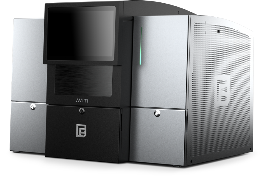
Short read sequencing on the Element Biosciences AVITI
The Element Biosciences AVITI is a recently purchased instrument that is cutting edge in the short read market. The AVITI is a mid sized sequencer, so flow cells provide ~1 billion reads/run or less. This makes it ideal for rapid turnaround times on projects because we are not waiting long periods of time to fill very large flow cells. Typically sequencing runs can be done from library receipt to data delivery in a matter of days. This instrument provides reads that are mostly in the Q40 range rather than the historically standard Q30 output from other competing sequencers, providing the user with exceptional data quality from their samples. The AVITI is also more tolerant of GC composition than standard sequencing. This allows for better sequencing overall as coverage will be more uniform across templates, especially difficult templates. We have seen this specifically in 10x Genomics libraries where more genes per cell are actually seen vs. Illumina sequencing. In addition to these differences, we have also seen that the AVITI is able to sequence through difficult regions (like homopolymer stretches) and then continue to provide usable sequence afterward. The AVITI is also able to sequence using larger insert sizes which may provide benefits in mapping data (for an example, check out the Google Deep Variant paper here). We have sequenced successfully in our lab with 1000bp inserts so far. We do not yet know where the upper limit is.
For further information please click here.
Long read sequencing with Oxford Nanopore Technologies (ONT)
Oxford Nanopore Technologies (ONT) technology provides multiple sequencing options provide for the ability to answer multiple questions. The P2Solo instrument we just recently received is able to run two PromethION flow cells at once. Each flow cell is sufficient to sequence one human genome. Smaller projects can be barcoded and combined into a single run. The instrument will sequence the DNA and also can obtain methylation state of the DNA at the same time. Because it is based on a pore rather than an enzymatic method, the ONT devices are the only systems on the market that can directly read RNA. The ONT system also has the unique ability to allow for sequencing of mostly regions of interest and throw out reads from areas outside the targeted regions. This mode is called adaptive sampling. A bed file is created with some buffer areas around your target regions. This bed file informs the sequencer which sequences are targeted. The sequencer reads the sequences as they pass through the pores in real time and after around 400-500 bases are read, if they don’t match the target regions, the sequencer reverses polarity of that one pore and ejects the DNA strand, making that pore available again for a different strand of DNA to enter. In this way, target regions can be enriched during the run without any special captures or amplification approaches.
Single cell sequencing with the 10x X Controller
The 10x X Controller is used for single cell analysis. Researchers are using this to evaluate cell types from different organs. Our controller can be used for all 10x protocols except spatial at this time. The DNA Sequencing core works hand in hand with the HSC Flow Cytometry core and HSC Bioinformatics core to provide a sample to data experience for our users.
Sanger sequencing on the Applied Biosystems 3730xl
The ABI 3730xl is the workhorse that has been used by industry for many years now and still continues to put out quality data for researchers here on campus and the surrounding areas. Turn around time on samples can be as quick as the same day.
In addition to the above, the DNA Sequencing core is always interested in helping with experiments that may not fit into the standard box envisioned by manufacturers of instrumentation and as such is always willing to discuss and help execute plans for novel and exciting additions to the technologies and techniques already in use in our core. Feel free to some talk to us about your project.
Service Rates
RNA Sequencing (poly A) Information
RNA Sequencing (poly A) is currently brand new and we are targeting batches of 16 samples. Lower numbers will increase the cost/sample as we need to fill the flow cell in order to cover costs. Please contact Derek with questions about projects and we can talk it through. At 16 samples, costs should be $250/transcriptome at approximately 40 million reads each.
Plasmid sequencing submission instructions can be found here.
Requesting Services
Existing users may login directly to the Resource Scheduling System to schedule or order services. This system is cores-wide and uses University of Utah uNID authentication.
Sanger Sample Submission Guidelines
PCR Products
- Quantitate using a gel (nanodrop isn’t reliable with PCR products)
- For small templates use 50ng PCR product (please clean it up first as left over primers can mess up sequencing) and 4 picomoles of primer (4uL of a 1uM aliquot)
- For larger templates, it is recommended to go high as 150ng (for approx. 2kb). Otherwise somewhere between the minimum and this will likely work. Use 4 picomoles of primer (4uL of a 1uM aliquot).
- Add molecular grade water to a total volume of 10uL
Plasmids
- For small samples (2-6kb) 600ng of product and 4 picomoles of primer (4uL of a 1uM aliquot) is recommended.
- For larger samples use more template/product (7-8kb: 700-800ng recommended, 9-10kb: 900-1000ng recommended) with 4 picomoles of primer (4uL of a 1uM aliquot).
- Add molecular grade water to a total volume of 10uL
Labeling your samples

Retrieving your data
The data is available the same night the run completes. The plate status, however, won't change until it has been reviewed by our staff. To find your data before we mark it as completed:
- Login to the Resource system
- Go to the "Sequencing Order Manager" (Located in the "DNA Sequencing" core)
- Change the View Mode from "Viewing: Completed" to "Viewing: Processing"
- Locate your order in the list and click on the "Detail" button with the magnifying glass
- Change the active tab (Green) from "Overview" to "Results". From the results tab you can download the whole dataset or the files for individual samples.
Once we have checked the plate and determined which samples we feel we need to redo your order will be marked as completed. To retrieve data from completed orders:
- Login to the Resource system
- Go to the "Sequencing Order Manager" (Located in the "DNA Sequencing" core)
- From the "Viewing: Completed" View mode you will see completed orders
- Locate your order in the list and click on the "Detail" button with the magnifying glass
- Change the active tab (Green) from "Overview" to "Results". From the results tab you can download the whole dataset or the files for individual samples
Install Instructions Here
Contact Us
Hours of Operation
9:00 am to 5:00 pm
Monday - Friday
Mailing Address
DNA Sequencing
30 N 2030 E, Rm 115
Salt Lake City, UT 84112
Recent Publications
- Balakrishnan, B., R. Altassan, R. Budhraja, W. Liou, A. Lupo, S. Bryant, A. Mankouski, S. Radenkovic, G.J. Preston, A. Pandey, S. Boudina, T. Kozicz, E. Morava-Kozicz and K. Lai (2023). AAV-based gene therapy prevents and halts the progression of dilated cardiomyopathy in a mouse model of phosphoglucomutase 1 deficiency (PGM1-CDG). Transl Res 257: 1-14.10.1016/j.trsl.2023.01.004
- Demir, M., L. P. Russelburg, W. J. Lin, C. H. Trasvina-Arenas, B. Huang, P. K. Yuen, M. P. Horvath and S. S. David (2023). Structural snapshots of base excision by the cancer- associated variant MutY N146S reveal a retaining mechanism. Nucleic Acids Res 51(3): 1034-1049.10.1093/nar/gkac1246
- Flack, C. E. and J. S. Parkinson (2022). Structural signatures of Escherichia coli chemoreceptor signaling states revealed by cellular crosslinking. Proc Natl Acad Sci U S A 119(28): e2204161119.10.1073/pnas.2204161119
- Fleming, A. M. and C. J. Burrows (2023). Nanopore sequencing for N1-methylpseudouridine in RNA reveals sequence-dependent discrimination of the modified nucleotide triphosphate during transcription. Nucleic Acids Res 51(4): 1914-1926.10.1093/nar/gkad044
- Fleming, A. M., R. Tran, C. A. Omaga, S. A. Howpay Manage, C. J. Burrows and J. C. Conboy (2022). Second Harmonic Generation Interrogation of the Endonuclease APE1 Binding Interaction with G-Quadruplex DNA. Anal Chem 94(43): 15027-15032.10.1021/acs.analchem.2c02951
- Gerstner, C. D., M. Reed, T. M. Dahl, G. Ying, J. M. Frederick and W. Baehr (2022). Arf-like Protein 2 (ARL2) Controls Microtubule Neogenesis during Early Postnatal Photoreceptor Development. Cells 12(1).10.3390/cells12010147
- Giglio, M. L., P. F. Salcedo, M. Watkins and B. Olivera (2023). Insights into a putative polychaete- gastropod symbiosis from a newly identified annelid worm that predates upon Conus ermineus eggs. Contributions to Zoology 92(2): 97-111.https://doi.org/10.1163/18759866-bja10038
- Hackney, C. M., P. F. Salcedo, E. Mueller, L. D. Kjelgaard, M. Watkins, L. G. Zachariassen, J. R. McArthur, D. J. Adams, A. S. Kristensen, B. Olivera, R. K. Finol-Urdaneta, H. Safavi-Hemami, J. P. Morth and L. Ellgaard (2022). Identification of a sensory neuron Cav2.3 inhibitor within a new superfamily of macro- conotoxins. bioRxiv: 2022.2007.2004.498665.10.1101/2022.07.04.498665
- Happ, J. T., C. D. Arveseth, J. Bruystens, D. Bertinetti, I. B. Nelson, C. Olivieri, J. Zhang, D. S. Hedeen, J. F. Zhu, J. L. Capener, J. W. Brockel, L. Vu, C. C. King, V. L. Ruiz-Perez, X. Ge, G. Veglia, F. W. Herberg, S. S. Taylor and B. R. Myers (2022). A PKA inhibitor motif within SMOOTHENED controls Hedgehog signal transduction. Nat Struct Mol Biol 29(10): 990-999.10.1038/s41594-022-00838-z
- Howpay Manage, S. A., A. M. Fleming, H. N. Chen and C. J. Burrows (2022). Cysteine Oxidation to Sulfenic Acid in APE1 Aids G-Quadruplex Binding While Compromising DNA Repair. ACS Chem Biol 17(9): 2583-2594.10.1021/acschembio.2c00511
- Howpay Manage, S. A., J. Zhu, A. M. Fleming and C. J. Burrows (2023). Promoters vs. telomeres: AP- endonuclease 1 interactions with abasic sites in G-quadruplex folds depend on topology. RSC Chem Biol 4(4): 261-270.10.1039/d2cb00233g
- Leng, A. M., K. S. Radmall, P. K. Shukla and M. B. Chandrasekharan (2022). Quantitative Assessment of Histone H2B Monoubiquitination in Yeast Using Immunoblotting. Methods Protoc 5(5).10.3390/mps5050074
- McKnite, A., H. S. Kim, J. Silva and J. L. Christian (2023). Lack of evidence that fibrillin1 regulates bone morphogenetic protein 4 activity in kidney or lung. Dev Dyn 252(6): 761-769.10.1002/dvdy.578 37
- Radmall, K. S., P. K. Shukla and M. B. Chandrasekharan (2023). A system for <em>in vivo</em> evaluation of protein ubiquitination dynamics using deubiquitinase-deficient strains. bioRxiv: 2023.2006.2018.545485.10.1101/2023.06.18.545485
- Reed, M., K. I. Takemaru, G. Ying, J. M. Frederick and W. Baehr (2022). Deletion of CEP164 in mouse photoreceptors post-ciliogenesis interrupts ciliary intraflagellar transport (IFT). PLoS Genet 18(9): e1010154.10.1371/journal.pgen.1010154
- Scoles, D. R., M. Gandelman, S. Paul, T. Dexheimer, W. Dansithong, K. P. Figueroa, L. T. Pflieger, S. Redlin, S. C. Kales, H. Sun, D. Maloney, R. Damoiseaux, M. J. Henderson, A. Simeonov, A. Jadhav and S. M. Pulst (2022). "A quantitative high-throughput screen identifies compounds that lower expression of the SCA2-and ALS-associated gene ATXN2." J Biol Chem 298(8): 102228.10.1016/j.jbc.2022.102228
- Simeone, C. A., J. L. Wilkerson, A. M. Poss, J. A. Banks, J. V. Varre, J. L. Guevara, E. J. Hernandez, B. Gorsi, D. L. Atkinson, T. Turapov, S. G. Frodsham, J. C. F. Morales, K. O'Neil, B. Moore, M. Yandell, S. A. Summers, A. S. Krolewski, W. L. Holland and M. G. Pezzolesi (2022). "A dominant negative ADIPOQ mutation in a diabetic family with renal disease, hypoadiponectinemia, and hyperceramidemia." NPJ Genom Med 7(1): 43.10.1038/s41525-022-00314-z
- Shukla, P. K., J. E. Bissell, S. Kumar, S. Pokhrel, S. Palani, K. S. Radmall, O. Obidi, T. J. Parnell, J. Brasch, D. C. Shrieve and M. B. Chandrasekharan (2023). Structure and functional determinants of Rad6- Bre1 subunits in the histone H2B ubiquitin-conjugating complex. Nucleic Acids Res 51(5): 2117- 2136.10.1093/nar/gkad012
- Shukla, P. K., K. S. Radmall and M. B. Chandrasekharan (2023). Rapid purification of rabbit immunoglobulins using a single-step, negative-selection chromatography. Protein Expr Purif 207: 106270.10.1016/j.pep.2023.106270
- Shukla, P. K., D. Sinha, A. M. Leng, J. E. Bissell, S. Thatipamula, R. Ganguly, K. S. Radmall, J. J. Skalicky, D. C. Shrieve and M. B. Chandrasekharan (2022). Mutations of Rad6 E2 ubiquitin-conjugating enzymes at alanine-126 in helix-3 affect ubiquitination activity and decrease enzyme stability. J Biol Chem 298(11): 102524.10.1016/j.jbc.2022.102524
- Utzman, P. H., V. P. Mays, B. C. Miller, M. C. Fairbanks, W. J. Brazelton and M. P. Horvath (2023). Metagenome mining and functional analysis reveal oxidized guanine DNA repair at the Lost City Hydrothermal Field. bioRxiv: 2023.2004.2005.535768.10.1101/2023.04.05.535768
- Walker, M. F., J. Zhang, W. Steiner, P.-I. Ku, J.-F. Zhu, Z. Michaelson, Y.-C. Yen, A. B. Long, M. J. Casey, A. Poddar, I. B. Nelson, C. D. Arveseth, F. Nagel, R. Clough, S. LaPotin, K. M. Kwan, S. Schulz, R. A. Stewart, J. J. G. Tesmer, T. Caspary, R. Subramanian, X. Ge and B. R. Myers (2023). GRK2 Kinases in the Primary Cilium Initiate SMOOTHENED-PKA Signaling in the Hedgehog Cascade. bioRxiv: 2023.2005.2010.540226.10.1101/2023.05.10.540226
- Workalemahu, T., C. Avery, S. Lopez, N. R. Blue, A. Wallace, A. R. Quinlan, H. Coon, D. Warner, M. W. Varner, D. W. Branch, L. B. Jorde and R. M. Silver (2023). Whole-genome sequencing analysis in families with recurrent pregnancy loss: A pilot study. PLoS One 18(2): e0281934.10.1371/journal.pone.0281934
Citing Our Facility
Acknowledgments
We would like to thank you for acknowledging the our facility. This recognition allows us to highlight the impact of your work and demonstrates the important contributions of our facility makes to research across the University of Utah. The recognition our core receives from your acknowledgments also aids in receiving grants and further funding for equipment and services we can provide to our users.
Self-Run Services / Instrumentation Usage:
In published papers that used instruments at our facility and notably involved staff members please use the following format:
We acknowledge (facility name) at the University of Utah for use of equipment (insert instrument/service details here), and thank (insert any notable staff member – if desired) for their assistance.
Assisted Services:
In published papers where a staff member assisted you in addition to the requested services please use the following format:
We acknowledge (facility name) at the University of Utah for use of equipment (insert instrument/service details here), and thank (insert staff member-required) for their assistance in (service provided).
Collaboration:
For publications resulting from collaborations that assisted with the methodologies, planning process and execution of your experiment in addition to equipment usage we require Co-author attribution on your publication for our facility and any staff members who provided substantial contributions to the originating project.


