Preclinical Imaging Core Facility
The Preclinical Imaging Core Facility was established in 2008 by Dr. Edward Hsu with the goal of providing advanced imaging instrumentation and expertise for small animal research at the University of Utah. The Preclinical (formerly Small Animal) Imaging Facility extends the benefits of modern diagnostic medical imaging technologies to the studies of anatomy and physiology in small animals. The facility features state-of-the-art MRI, CT, PET, SPECT, and MSOT systems. All instruments are equipped with supporting and monitoring hardware that allows a wide variety of imaging experiments, including longitudinal studies, to be performed on live animals and specimens. Imaging scientists, full-time imaging personnel, and animal support technicians are available for technical consultation and experimental assistance.
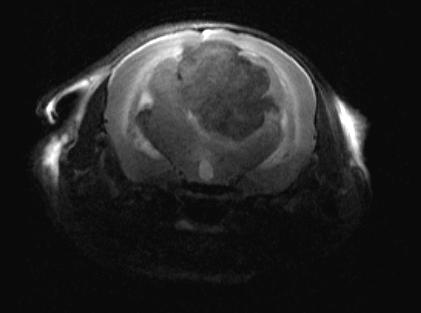
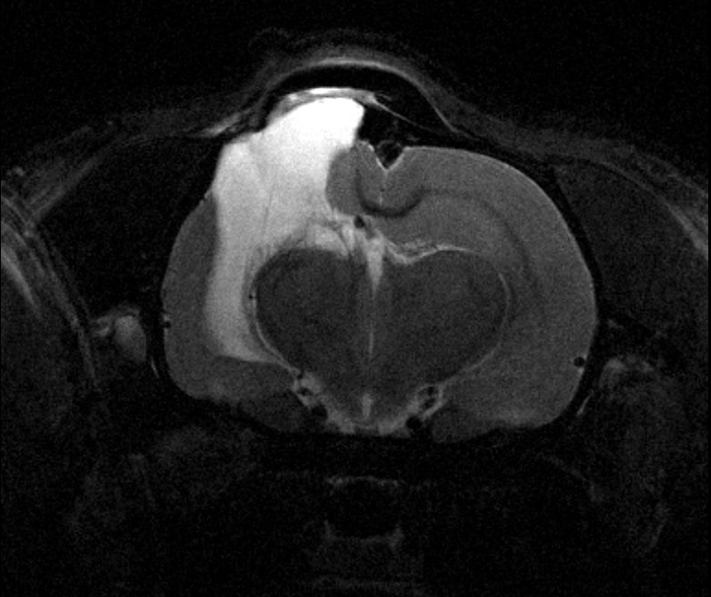
The Bruker Biospec 7 Tesla Preclinical MRI system is optimal for most MRI and NMR spectroscopy studies. System capabilities include DTI studies, In-vivo gated CINE scans, TOF vasculature studies, T2-Weighted, T1-Weighted anatomy scans [BW1], and neuroimaging studies.
The Preclinical Imaging facility has two preclinical computed tomography (CT) scanners for use with live animals or specimens. Uses include embryonic and adult specimen anatomy, bone mineral density, bone mineral content, angiogenesis index, and fossil and geological sample assessment. The Inveon PET/SPECT/CT Trimodality Preclinical system is an excellent choice for multimodality scans. Example projects include 10-micron resolution mice skull scans, large FOV heart scans, FDG cancer studies, and vasculature studies. A gamma counter is also available for nuclear medicine tracer calibration and quantification.
Our facility houses an Ithera MSOT optoacuostic tomography system that allows for in-vivo blood oxygenation and molecular tracer imaging in rats and mice.
Rates
Available Services
| Rate Name | Internal (UoU) | Academic | Commercial | |
| MRI | Live Animal | $ 315.00 / hour | $ 486.00 / hour | Available on request |
| Specimen (4 hour minimum) | $ 90.00 / hour | $ 139.00 / hour | ||
| Processing and Analysis | $ 100.00 / hour | $ 154.00 / hour | ||
| CT | Staff Service | $ 315.00 / hour | $ 486.00 / hour | |
| Processing and Analysis | $ 100.00 / hour | $ 154.00 / hour | ||
| PET/SPECT | Staff Service | $ 250.00 / hour | $ 385.00 / hour | |
| Self Service - Training Required | $ 150.00 / hour | $ 235.00 / hour | ||
| Processing and Analysis | $ 100.00 / hour | $ 154.00 / hour | ||
| MSOT | Self Service - Training Required | $ 52.50 / hour | $ 81.00 / hour | |
| Staff Service | $ 100.00 / hour | $ 154.50 / hour |
MRI
Our Preclinical Imaging Facility is equipped with a state-of-the-art 7 Tesla Bruker BioSpec MRI scanner with a 30 cm wide cylindrical bore for NMR imaging and spectroscopy applications. Typical preclinical models include rabbits, rats, and mice. Some of the features of our MRI scanner are:
- 7.05 Tesla magnetic field at magnet isocentre
- Actively shielded magnet (reduced stray field)
- 4 receiver channels and parallel imaging capabilities
- Scan gating using respiration or ECG signal
- 3 interchangeable gradient sets
- i.d. = 20 cm, max gradient = 200 mT/m (shielded) [For larger specimens and animals such as rabbits]
- i.d. = 12 cm, max gradient = 400 mT/m (shielded) [For medium sized specimens and animals such as rats and mice]
- i.d. = 6 cm, max gradient = 1000 mT/m (unshielded) [For mice applications only]
- Preemphasis unit for eddy current compensation
- Multi-nuclear NMR capability:1H, 31P, 19F, 13C
- Modular software development: pulse programs, methods, macros.
- 15 commercially available volume RF coils ideal for brain imaging (rats and mice) to whole body applications. { Bruker 1H rat brain surface coil , Bruker 1H mouse brain surface coil, Bruker 1H quadrature volume coil with i.d = 72 mm, Bruker 1H linear volume coil with i.d = 72 mm, Bruker 1H quadrature volume coil with i.d = 150 mm, M2M 1H quadrature volume coil with i.d = 90 mm, Rapid 1H quadrature volume coil with I.d = 35 mm and 50 mm}
Our facility also includes an Instrument Development Lab which primarily provides infrastructure for the construction of custom RF coils. These are often necessary to optimize the data quality for a given MRI application. We offer basic machining and 3D printing operations for making experimental apparatus such as scanning platforms and stereotaxes.
Some of our MR imaging capabilities include but are not limited to:
- High-resolution anatomic imaging of small animals or biological specimens
- Diffusion-weighted or diffusion tensor imaging
- Relaxometry (T1, T2, T2*) mapping
- Perfusion MRI
- Functional MRI
- MR angiography
- Cardiac MRI
- NMR spectroscopy (localized and non-localized)
- Chemical shift imaging
- Parallel imaging techniques
Examples of current applications can be viewed in our Gallery section.
CT
Our facility also houses the Inveon Multimodality System is a versatile platform for laboratory animal CT, SPECT, and PET studies on a single integrated gantry. The two scanners in the system can operate independently or as a multimodality system under the control of a single workstation.
Some features of the multi modal system are:
- Automatic transition between modes and seamless coordination of CT, SPECT, and PET data
System can configured as an ultra-high resolution preclinical CT scanner; - High-resolution, high-sensitivity preclinical SPECT scanner
- As a dual modality preclinical SPECT/CT scanner.
- CT component of the device has a large area 165 mm X-ray camera. It incorporates a high-resolution, low-noise, 14-bit x-ray imaging detector with 4064 x 4064 pixels.
The Inveon 2-Head SPECT Module is designed to efficiently detect gamma rays ranging in energy from 30 keV to 250 keV, the SPECT system is ideal for use with most single photon-emitting radionuclides. The two large area detectors (15 cm X 15 cm) support whole animal studies as well as high magnification studies.
The system includes two Inveon Research Workplace workstations for multimodality image review, fusion, and analysis which CT, PET, SPECT, and MR data in DICOM and Siemens Inveon CT, PET, and SPECT formats, as well as raw data import.
iThera MSOT inVision- 256 TF
This is a hybrid photoacoustic imaging system for visualizing optical contrast at high resolution (down to 120 um) in deep tissue (upto 3 cm) and displaying the images in real time. The technology relies on the photoacoustic effect – the conversion of light energy into ultrasound signals. Tissues undergo thermoelastic expansion after they are illuminated by a pulsed laser, which generates ultrasound waves that are detected by a 270° array of ultrasound detectors. The whole mouse body can be scanned by moving the mouse through the detector ring.
The system allows study of versatile physiological processes at cellular and molecular level. Contrast can be endogenous (deoxy hemoglobin vs oxy hemoglobin, melanin, collagen, lipids etc) or from exogenous agents such as fluorescent dyes or nanoparticles. Both mice and rats can be accommodated.
Technical Specifications:
- Multispectral imaging capability
- Laser wavelength range: 680-980 nm/660-1300 nm
- Peak pulse energy: 100 mJ
- 5 MHz detector with 256 elements covering 270°
- 120 µm in-plane resolution
- 100 ms temporal resolution
- 20 x 20 mm cross-section which can increase to 40 x 40 mm.
- 2D cross sectional images obtained in real time. A stack of cross sectional images can be rendered into a 3D image offline.
Examples of applications:
- Perfusion MSOT
- Oxygenation MSOT
- In vivo biodistribution of probes and therapies
- Molecular imaging of diseases (cancer, inflammation, organ dysfunction, metabolic disorders, fibrosis, neurological disorders etc)
Siemens PET
The Siemens Inveon PET-CT system allows nuclear medical imaging that produces a three-dimensional image of functional processes in the body. The system detects pairs of gamma rays emitted indirectly by a positron-emitting radionuclide (tracer), which is introduced into the body on a biologically active molecule or nano/microparticle. The system has 4,064 × 4,064 detectors, a FOV greater than 10 × 10 cm, a spatial resolution of 15 micron isotropic voxels, and can scan an entire mouse in less than 1 minute. Siemens Inveon PET/CT Multimodality System allows PET and CT studies on a single integrated gantry.
In preclinical studies, PET imaging can be used for noninvasive detection and investigation of diseases, assessment of biodistribution and pharmacokinetics of small molecule, biologics and nanoparticles in small animal models. Because of its superior quantification and sensitivity, PET has been applied in understanding of disease mechanisms, diagnosis, treatment planning, and therapeutic efficacy monitoring in cancer and other diseases.
Requesting Services
Existing users may login directly to the Resource Scheduling System to schedule or order services. This system is cores-wide and uses University of Utah uNID authentication.


Hours of Operation
9:00 am to 5:00 pm
Monday - Friday
Location
Sorenson Molecular Biotechnology Building
36 S Wasatch Dr
Salt Lake City, UT 84112
Major Citations
- Robinson, J. P., Vanbrocklin, M. W., McKinney, A. J., Gach, H. M. & Holmen, S. L. Akt signaling is required for glioblastoma maintenance in vivo. Am. J. Cancer Res. 1, 155–167 (2011).
- Abdullah OM, Darkos SG, Diakos NA, Wever-Pinzon O, Kfoury AG, Stehlik J, Selzman CH, Reid BB,
Brunisholz K, Verma DR, Myrick C, Sachse FB, Li DY, Hsu EW. (2014). Characterization of diffuse
fibrosis in the failing human heart via diffusion tensor imaging and quantitative histological validation. NMR Biomed; 27(11):1378-86. PMID: 25200106. - Welsh C, Di Bella E, Hsu E. (2015) Higher-order motion-compensation for in vivo cardiac diffusion
tensor imaging in rats. IEEE Trans Med Imaging; 34(9): 1843-53. PMID: 25775486. - Merchant SS, Gomez AD, Morgan JL, Hsu EW. (2016). Parametric modeling of the mouse left
ventricular myocardial fiber structure. Ann Biomed Eng. 44(9):2661-73. PMID: 26942586. - Pritz, M. B., Ziegler, L. C., Thompson, T. N. & Hsu, E. W. Magnetic resonance diffusion tensor tractography of a midbrain auditory circuit in Alligator. Neurosci. Lett. 738, 135251 (2020).
Recent Mentions
- Boer, E. F., E. T. Maclary and M. D. Shapiro (2021). Complex genetic architecture of three-dimensional craniofacial shape variation in domestic pigeons. Evol Dev 23(6): 477-495.10.1111/ede.12395
- Epperson, R. T., B. M. Isaacson, D. L. Rothberg, R. E. Olsen, B. Kawaguchi, J. M. Maxwell, M. Dickerson, P. F. Pasquina, J. Shero and D. L. Williams (2021). Developing a combat-relevant translatable large animal model of heterotopic ossification. Bone Rep 15: 101127.10.1016/j.bonr.2021.101127
- Jeong, K. E., S. Y. Lee, S. K. Yeom, N. Carlson, L. M. Shah, J. Rose and E. K. Jeong (2022). Ultrahigh-b diffusion-weighted imaging for quantitative evaluation of myelination in shiverer mouse spinal cord. Magn Reson Med 87(1): 179-192.10.1002/mrm.28978
- Melstrom, K. M., A. H. Turner and R. B. Irmis (2022). Reevaluation of the cranial osteology and phylogenetic position of the early crocodyliform Eopneumatosuchus colberti, with an emphasis on its endocranial anatomy. Anat Rec (Hoboken) 305(10): 2557-2582.10.1002/ar.24777
- Merchant, S., S. Yeoh, M. A. Mahan and E. W. Hsu (2022). Simultaneous Quantification of Anisotropic Microcirculation and Microstructure in Peripheral Nerve. J Clin Med 11(11).10.3390/jcm11113036
- Patel, M., F. Savvopoulos, C. C. Berggren, L. Aslanidou, L. H. Timmins, R. de Silva, R. M. Pedrigi and R. Krams (2021). Considerations for analysis of endothelial shear stress and strain in FSI models of atherosclerosis. J Biomech 128: 110720.10.1016/j.jbiomech.2021.110720
- Smith, K. A., S. S. Merchant, E. W. Hsu and L. H. Timmins (2021). Effect of Subject-Specific, Spatially Reduced, and Idealized Boundary Conditions on the Predicted Hemodynamic Environment in the Murine Aorta. Ann Biomed Eng 49(12): 3255-3266.10.1007/s10439-021-02851-7
- Welsh, P. J., C. G. Collier, H. M. Clement, M. N. Vakula and J. B. Mason (2021). Cranial Cruciate Ligament Desmotomies in Sheep Resulting in Peroneus Tertius Injury. Case Rep Vet Med 2021: 2628791.10.1155/2021/2628791
- Yeoh, S., W. S. Warner, S. S. Merchant, E. W. Hsu, D. V. Agoston and M. A. Mahan (2022). Incorporating Blood Flow in Nerve Injury and Regeneration Assessment. Front Surg 9: 862478.10.3389/fsurg.2022.862478
Citing Our Facility
Acknowledgments
We would like to thank you for acknowledging the our facility. This recognition allows us to highlight the impact of your work and demonstrates the important contributions of our facility makes to research across the University of Utah. The recognition our core receives from your acknowledgments also aids in receiving grants and further funding for equipment and services we can provide to our users.
Self-Run Services / Instrumentation Usage:
In published papers that used instruments at our facility and notably involved staff members please use the following format:
We acknowledge (facility name) at the University of Utah for use of equipment (insert instrument/service details here), and thank (insert any notable staff member – if desired) for their assistance.
Assisted Services:
In published papers where a staff member assisted you in addition to the requested services please use the following format:
We acknowledge (facility name) at the University of Utah for use of equipment (insert instrument/service details here), and thank (insert staff member-required) for their assistance in (service provided).
Collaboration:
For publications resulting from collaborations that assisted with the methodologies, planning process and execution of your experiment in addition to equipment usage we require Co-author attribution on your publication for our facility and any staff members who provided substantial contributions to the originating project.